Skeleton Back Bones Diagram - Medical Education Chart of Biology for Human Skeleton ... : *the origin, insertion, and belly.* a muscle's origin is where a tendon attaches it to the *less* movable bone.
Skeleton Back Bones Diagram - Medical Education Chart of Biology for Human Skeleton ... : *the origin, insertion, and belly.* a muscle's origin is where a tendon attaches it to the *less* movable bone.. As you can see in the above diagram, there are around 24 labels, and forgetting one of them is quite possible. The longest and the strongest bone in the human skeletal system as you can observe in the labeled skeleton diagram of the human body. Spinal vertebrae bone spine vertebra toracica spinal cord spine structure back diagram spine sections spinal cord vertebrae spinal structure health diagram. Posted in bones, diagrams | tagged body skeleton, human skeletal anatomy, human skeleton, human. A lateral view skeletal diagram offers a side view of the human skeleton.
Your lower back contains 5 vertebral bones stacked above each other with intervertebral discs in between. Vertebrae, bones, joints, ligaments, muscles, muscular system, fascia, arteries, veins, nerves and various adjacent organs. These bones are connected at the back with specialized joints. See lumbar spine anatomy diagram stock video clips. Vertebrae separated by intervertebral discs.
axial skeleton - Wikidata from upload.wikimedia.org As you can see in the above diagram, there are around 24 labels, and forgetting one of them is quite possible. The longest and the strongest bone in the human skeletal system as you can observe in the labeled skeleton diagram of the human body. See lumbar spine anatomy diagram stock video clips. Front view of muscles , back view of muscles , organs , nervous system This diagram depicts back skeletal anatomy with parts and labels. The cranial bones compose the top and back of the skull and enclose the brain. The bones of the appendicular skeleton (the limbs and girdles) append to the axial skeleton. Many muscles that move the trunk and legs, such as our abdominal muscles, attach to the hip bones.
The bones of the skeletal system act as attachment points for the skeletal muscles of the body.
Using this atlas of human anatomy of the spine and back. Bones of the body 12 photos of the bones of the body bones of the body anatomy, bones of the body blank diagram, bones of the body jingle, bones of the body quiz game, number of bones. These bones are connected at the back with specialized joints. The cranial bones compose the top and back of the skull and enclose the brain. Almost every skeletal muscle works by pulling two or more bones either closer together or further apart. The arch is made of two supporting pedicles and two laminae (fig. It's made up of the bones that form the vertical axis of the body, such as the bones of the head, neck, chest, and spine. The back muscles are skeletal muscles. Human back bone chart, find out more about human back bone chart. Related posts of human back bones diagram female pelvis bones images. Individual anatomical structures include 2: This article looks at the anatomy of the back, including bones, muscles, and nerves. They support bones, in this case, the vertebrae.
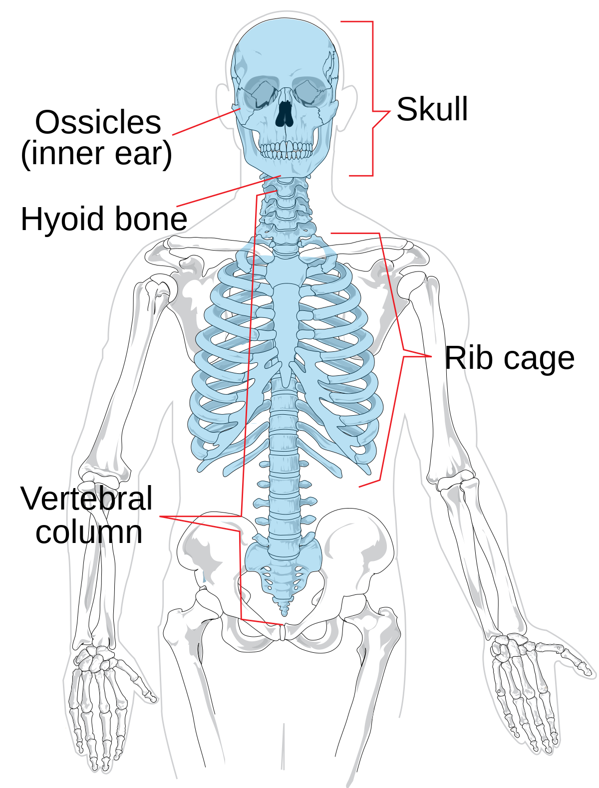
The red lines point individual bones and the names are writen in singular, the blue lines conect to group of bones and are in plural form. Human back bone chart, find out more about human back bone chart. Your lower back contains 5 vertebral bones stacked above each other with intervertebral discs in between. Female pelvis bones images 12 photos of the female pelvis bones images female human pelvis images, female pelvic bones images, bone, female human pelvis images, female pelvic bones images In addition, the broad hip bones provide protection to the delicate internal organs of the pelvis, such as the intestines, urinary bladder, and uterus.
al13nd2 from sites.google.com It runs down the centre of the body. Posted on june 17, 2016 by admin. On the back of each vertebra are bony projections that form the vertebral arch. The arch is made of two supporting pedicles and two laminae (fig. Spinal vertebrae bone spine vertebra toracica spinal cord spine structure back diagram spine sections spinal cord vertebrae spinal structure health diagram. Front view of muscles , back view of muscles , organs , nervous system It holds and protects the organs. The hollow spinal canal contains the spinal cord, fat, ligaments, and blood vessels.
This article looks at the anatomy of the back, including bones, muscles, and nerves.
The skeleton provides a framework for the body. Front view of muscles , back view of muscles , organs , nervous system This article looks at the anatomy of the back, including bones, muscles, and nerves. The hollow spinal canal contains the spinal cord, fat, ligaments, and blood vessels. The longest and the strongest bone in the human skeletal system as you can observe in the labeled skeleton diagram of the human body. Related posts of human back bones diagram female pelvis bones images. Each typical vertebra consists of a body, an arch and three processes that stem from. A posterior view skeletal diagram provides a back view of the human skeleton. Bones of the body 12 photos of the bones of the body bones of the body anatomy, bones of the body blank diagram, bones of the body jingle, bones of the body quiz game, number of bones. It runs down the centre of the body. The cranial bones compose the top and back of the skull and enclose the brain. The vertebral column, also known as the backbone or spine, is part of the axial skeleton.the vertebral column is the defining characteristic of a vertebrate in which the notochord (a flexible rod of uniform composition) found in all chordates has been replaced by a segmented series of bone: Vertebrae are the structural constituents of the spine.there are 33 vertebrae in total;
On the back of each vertebra are bony projections that form the vertebral arch. A posterior view skeletal diagram provides a back view of the human skeleton. The ilium is the big bone of the hip, the ischium is the bone on which one sits and the pubis forms the lower frontal hip bone as seen in the diagram. These bones are connected at the back with specialized joints. The hollow spinal canal contains the spinal cord, fat, ligaments, and blood vessels.
30 Label The Skeleton Worksheet Pdf - Labels For Your Ideas from lh6.googleusercontent.com The human skeleton, like that of other vertebrates, consists of two principal subdivisions, each with origins distinct from the others and each presenting certain individual features.these are (1) the axial, comprising the vertebral column—the spine—and much of the skull, and (2) the appendicular, to which the pelvic (hip) and pectoral (shoulder) girdles and the bones and cartilages of the. The ilium is the big bone of the hip, the ischium is the bone on which one sits and the pubis forms the lower frontal hip bone as seen in the diagram. Skull bones protect the brain and form an entrance to the body. The si joints are located on either side of the sacral spine and are situated deep in the. The bones of the skeletal system act as attachment points for the skeletal muscles of the body. Related posts of human back bones diagram female pelvis bones images. Your lower back contains 5 vertebral bones stacked above each other with intervertebral discs in between. It also covers some common conditions and injuries that can affect the back.
It also provides a structure for the interconnecting muscles.
Vertebrae, bones, joints, ligaments, muscles, muscular system, fascia, arteries, veins, nerves and various adjacent organs. The back muscles are skeletal muscles. Bones, discs, and joints in your lower back. The human skeleton, like that of other vertebrates, consists of two principal subdivisions, each with origins distinct from the others and each presenting certain individual features.these are (1) the axial, comprising the vertebral column—the spine—and much of the skull, and (2) the appendicular, to which the pelvic (hip) and pectoral (shoulder) girdles and the bones and cartilages of the. Related posts of human back bones diagram female pelvis bones images. The arch is made of two supporting pedicles and two laminae (fig. Each typical vertebra consists of a body, an arch and three processes that stem from. Front view of muscles , back view of muscles , organs , nervous system The red lines point individual bones and the names are writen in singular, the blue lines conect to group of bones and are in plural form. They support bones, in this case, the vertebrae. Vertebrae are the structural constituents of the spine.there are 33 vertebrae in total; The cranial bones compose the top and back of the skull and enclose the brain. Head and neck test questions gross anatomy all cervical vertebra have a:
Skull bones protect the brain and form an entrance to the body back bones diagram. Your lower back contains 5 vertebral bones stacked above each other with intervertebral discs in between.


0 Komentar